2. The Treatments
There are cases when patients cause their recession by brushing too hard, but in most cases, the patient should not feel guilty regarding the cause of their problem. Recession is usually something to which the patient has been pre-disposed.
1. The Causes
The causes of Gingival Recession: Thin, Fragile Tissues vary and may be described as:
a. Prominent Roots of the Teeth
b. Muscle Attachments
c. Associated with Orthodontic Treatment
d. Trauma (physical damage) to the Gums
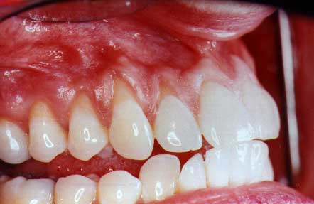
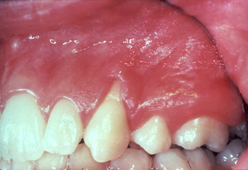
These lower teeth have advanced recession.The teeth themselves are almost "pushed" through the bone and gum. The lack of thick gum tissue is due to these roots being so prominent.
When a tooth erupts into the mouth, it should be surrounded by a layer of bone. If the bone is of a normal thickness, then it causes the developing gum tissue (which will cover the bone) to develop as a thick, tough layer of tissue. Nature designed this tissue to be tough in order to withstand the forces of chewing food hitting the gums.
If the developing bone is thin or absent on one side of the tooth (usually the lip or cheek side) the gum tissue that develops will be thin and fragile and not be able to withstand the normal forces that are applied to it from chewing and proper brushing.
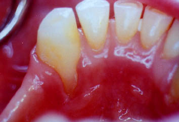
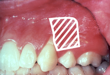
There is a muscle attachment at the base of this recession. This muscle attachment creates tension on the gum tissue and can cause recession to occur -- usually occuring on teeth which have prominent roots.
The mouth is a series of muscles which give you not only the ability to chew, but also to move your lips in ways such as "puckering up" your lips for a kiss. Some of these muscles originate under the lips and skin and have muscle attachments which insert into the gums -- usually not right where the gums meet the teeth.
If the attachments are right on the margin of where the gums meet the teeth, or if the attachments are very large, and especially if the roots of the teeth are prominent, there is the potential for recession.
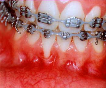
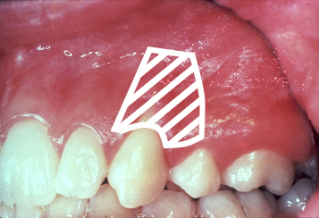
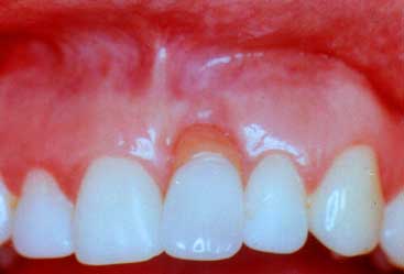
This recession is diagnosed during the course of her orthodontic treatment. The cause of the problem is the prominent roots due to the lack of heavy bone. The patient's jaw bone limits the orthodontist as to where he can move the teeth. The orthodontist's work is perfect!
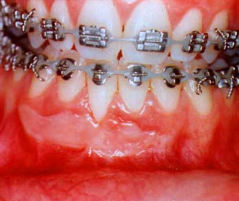
Patients undergoing orthodontic treatment may have recession treated before they begin their orthodontic treatment, during their orthodontic treatment or after they have completed their orthodontic treatment. The primary problem with recession that is associated with orthodontic treatment is that many times the patients do not have heavy, thick layers of bone over the roots of the teeth which are to be moved.
If the teeth are very crowded before orthodontic treatment, the only way to put the teeth into a good alignment may be if the roots of the teeth are made more prominent. The finest of orthodontic treatment can not overcome the lack of enough space in the jaw where the teeth could be moved without making the roots of the teeth more prominent. This is especially true when you consider how many parents do not wish for the orthodontist to extract teeth as part of the orthodontic treatment plan! There are also circumstances where recession will still occur even after some extractions were done.
Perfect orthodontic treatment can not overcome what nature did not adequately provide!!
The excessive wear of these teeth is related to her excessive and vigorous brushing with a brush that was "stiff", not "soft".
Trauma is "physical damage" to the gums where too much force is applied to the gums. The gums then "wear away" since they can't tolerate this excessive force. The two major examples of trauma are::
1. Toothbrushing Trauma.
This is where the patient either brushes too hard, too often or with too hard of a brush (only a "soft" bristled brush should be used).
2. Trauma from a Removable Partial Denture.
Patients who wear a removable partial denture risk recession to the gums usually on the tongue side of the lower teeth. If the partial denture fits against the gums too snugly, it will irritate the gums or "strip" the gums away from the teeth. A removable partial denture that has been worn for years or a new denture that "hurts" on the gums or "creates pressure" may increase the chances of getting recession.
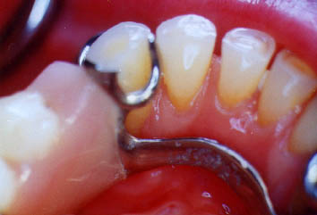
The tooth had its gums stripped due to the partial denture resting on the gums
This shows the partial denture as you are looking at the tongue side of the teeth. The partial denture puts pressure on the gums
The treatments for gingival recession vary depending on the amount of recession, the type, the cause and also the expected outcome of the treatment. Certain Periodontal Plastic Surgery procedures can create thick tissue which will stop further recession but in some cases these procedures may not be able to cover existing recession. The plastic surgery procedures are:
a. The Free Gingival Graft
b. The Lateral Graft
c. The Sub-Epithelial Connective Tissue Graft
a. The Free Gingival Graft
The Free Gingival Graft is a procedure where a thin piece of gum tissue is removed from the roof of the patient's mouth and transplanted to another site in order to create thick tough tissue or "attached gingiva".
In some cases, this type of gum graft can only prevent further recession, which is all that is necessary to keep the tooth or teeth healthy. In other cases, this type of gum graft can partially or almost completely cover previously existing recession.
The donor site (the roof of the mouth) will heal without damaging the underlying gums,bone or teeth. In some cases, a freeze-dried human tissue product can be used in order to avoid taking the tissue from the roof of the mouth.
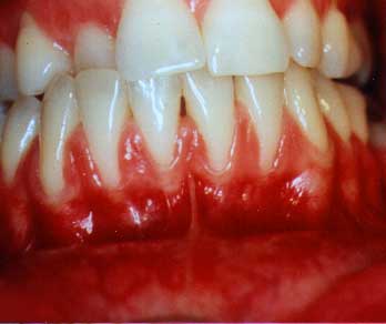
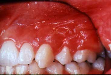
This recession is not only unsightly but is also infected since she can't clean these thin, fragile tissues properly.
After the gum graft has been done, you can see that the root has been partially covered. It is more difficult to cover roots if multiple teeth are involved
The lateral graft is also called a "pedicle" graft since the gum tissue is raised from the area immediately lateral, or adjacent, to the area of recession. In order to do this, there must be an abundant amount of thick, tough, "attached gingiva" lateral to this area of recession. This is frequently not the situation in many cases of recession.
This "pedicle" of gum tissue is then rotated over the area of recession in order to attempt to cover the recession, as well as create a thick band of "attached gingiva" which will prevent further recession. A limitation of this procedure is that there can be some recession occuring from where the "pedicle" was moved from.
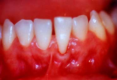
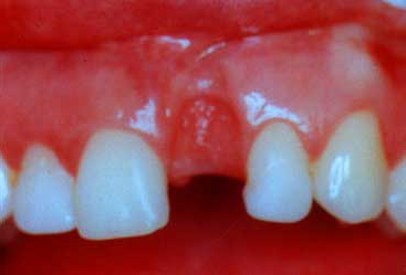
The patient is unhappy with this recession which is readily visible to her and others
The artist's drawing shows from where the "pedicle" flap will originate.
The artist's drawing shows how the "pedicle" flap will be rotated over in order to cover the recession.
The final healed result has created a good cosmetic result while also preventing further recession and disease
The sub-epithelial connective tissue graft is a procedure that is designed to:
1. Cover Recession (when a lateral "pedicle" graft can't be done)
2. Re-Construct Areas of Bone Loss or Bone Resporption
1. Cover Recession
When there is not enough thick tissue next to an area of recession and a lateral graft can't be done, the "Sub-Epi" graft can be done. This usually uses a piece of gum tissue taken from the roof of the mouth but from under the superficial, surface gum tissue. An option, in some cases, is to use a "man-made" membrane instead of taking any tissue from the roof of the mouth.
In the surgical procedure, the gum tissue or "man-made" membrane is placed over the root of the tooth. A flap of gum tissue is raised from the base of the recession in order to cover the gum tissue or membrane -- in order to provide a blood supply to the graft. This creates the potential to create healthy, tough tissue and also create a better cosmetic result.
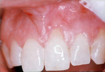
This tooth has severe recession that is very unsightly.
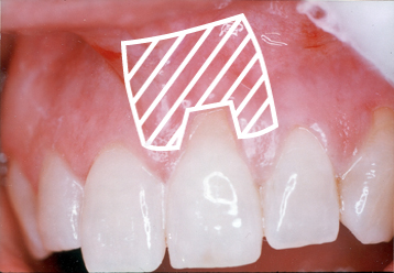
The "drawing" shows where a flap of tissue will be raised to cover the graft.
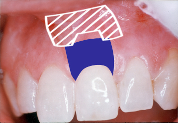
This drawing shows the gum tissue or the membrane (shaded blue for contrast) placed over the root.
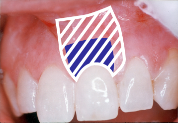
This drawing shows how the flap from the base of the recession is now covering the graft.
The healed result has covered the root and created a pleasing appearance.
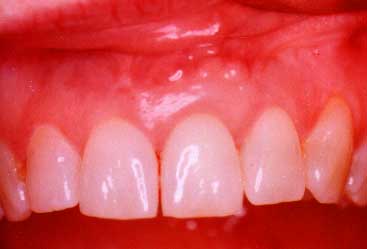
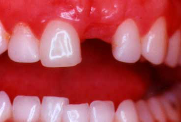
When some of the upper front teeth are removed (usually if a tooth fractured or a root canal failed), there may be the excessive loss of bone from where the tooth came out. This can leave an unsightly, or even ugly "depression" in the gum tissue. Too often, when a "dummy" tooth is replaced into the mouth, this "depression" requires the "dummy" or replacement tooth to be too long and unsightly. Before the replacement tooth is to be made, the "Sub-epi" graft should be done in order to reconstruct this deformed area. The result will be a more natural looking tooth and a happier smile.
This is a bridge with a very poor appearance. This long replacement tooth could have been avoided with a "Sub-epi" graft.
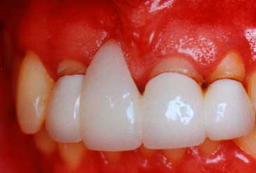
The "dummy"tooth on her partial denture has an ugly, discolored plastic extension protruding into the area of bone loss
With the tooth taken out, you can see how the plastic extension has "dug" into the gum tissue. A "Sub-Epi" graft will be placed under this depression in order to "plump-out" the gums.
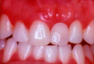
This shows the reconstructed area. The gum has been built-out as a result of the "Sub-epi" graft being placed under the gum to "bulk-out" this area.
This shows how her partial denture replacement tooth now looks with the ugly plastic extension removed.